
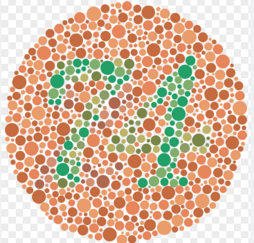
An Ishihara test to test colorblindness
Color is everywhere in our world. From the lush green of grass growing on a field to the rich deep blue of the ocean, every object we see has a color to it. However, not all people can see that vivid hue. Some are blind and lack the ability to see in general. Others are able to see, but their perception of color is lacking. Colorblindness affects a sizable portion of our population and is an interesting condition that plagues many people’s eyes.
Table of Contents:
- Origins and Sources
- Physical Issues
- Different Types
- Conclusion
Origins and Sources
Color blindness is usually a genetic and hereditary condition. Red/green or yellow/blue color blindness are passed down from parents on the X chromosome. This gene is recessive meaning that if one of the X chromosomes for a person has it and the other one (there are two total) does not, then the individual will not have the condition. This is the main reason why males (who have one x- chromosome and one y-chromosome) are more prone for this condition compared to females (two x-chromosomes) as both x-chromosomes need to have colorblindness as a gene for females whereas males only need one. The vast majority of people with a color vision deficiency have inherited their condition from their mother, who is normally a carrier of the gene but not color blind herself. The condition does not worsen as an individual ages; rather, it stays the same severity as from childhood. Overall there are an estimated 300 million people across the globe who have this condition, making it somewhat common.
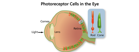
The Physical Issues
In the eye, there are two sets of light-sensitive nerve cells: rods and cones. Rod cells help with low-light level vision such as during the night whereas cone cells help distinguish color. There are 3 types of cone cells: red, blue, and green which are each sensitive to a different frequency. The brain combines the signals from all of the cells to form the color spectrum that we see. However, when one or more of the cone cell types has abnormal sequencing, color blindness occurs. In severe cases, one cone cell type may not exist at all and the person is called a dichromat.
Different types:
There are four main types of colorblindness. First is deuteranomaly, the most common type. It affects the green cone cells and causes red-green colorblindness. Another similar one is protanomaly which also causes red-green colorblindness, but this time affects the red cells. While they affect different cones, they end up looking very similar. However, in deuteranomaly, the greens become more reddish whereas in protanomaly the reds become more greenish. A third more rare type is called tritanomaly where the blue cones are affected causing blues to appear duller and muted. All of these are when the cones themselves are still present but are weak.
If the cones are missing, the spectrum of colors visible changes drastically. The suffix “-anomaly” means the cones are weak, but the suffix “-anopia” means the cones are missing. Deuteranopia is the most common with green cones missing, then protanopia and tritanopia. Lastly, the most rare form is monchromacy where an individual has only one cone or even no cones at all, making their entire world a grayscale. Only 1 in every 33,000 people have it thought making it much less common compared to the other forms.

Normal vision of a food stall

Deuteranopian vision of the same food stall
Conclusion:
Colorblindness affects a large percentage of the population. The color in our lives that we take for granted is invisible to some. Next time you look around at the purple flowers or the blue sky, take a minute and enjoy the spectrum of hue in the world.

https://shorturl.fm/yjk8O
Good day everyone, I wanted to share my thoughts on [url=https://10bet-online.com/]10Bet[/url] which I have been using for a couple months now. The platform is quite intuitive and the customer service team has been responsive to my inquiries. I would say its worth a look if your searching for something reliable. Best regards.